![]()
| Introduction
Major Arteries |
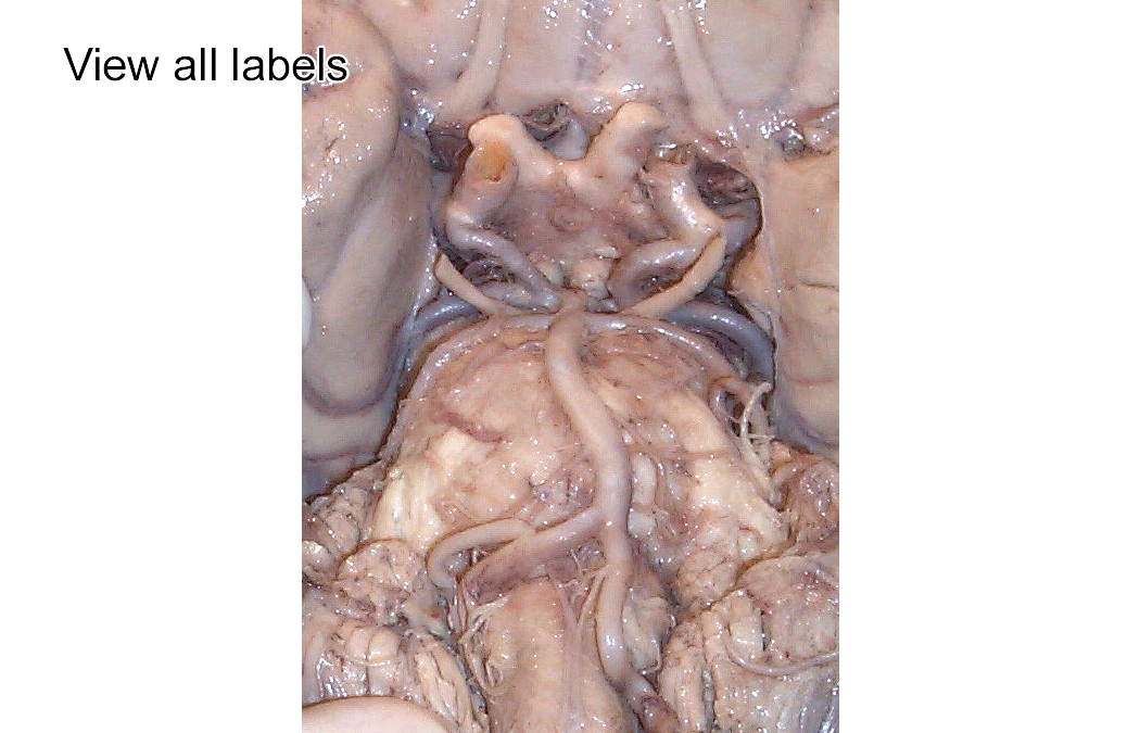
Click on hot spots in the image below to identify the following arteries. Click "View all labels" to see the labels; click on "Hide all labels" to return to the original image.
The blood supply of the brain comes from two primary sources: the internal carotid arteries, which supply blood to most of the telencephalon and diencephalon, and the vertebral arteries, which supply blood to the caudal third of the cortex and the entire brainstem and cerebellum. The two systems are connected by the posterior communicating artery which runs between the posterior and internal carotid arteries. The posterior cerebral, posterior communicating, internal carotid, anterior cerebral and anterior communicating arteries form an apparent circle of arteries that wrap around the pituitary stalk and optic nerve. This circle is called the Circle of Willis. The communicating arteries are supposed to supply an alternate path for blood flow in case of blockage of one of the major supply arteries. Since blood pressure is about the same on both sides of the communicating arteries, there may be limited blood flow through them. Over time, a slow, progressive occlusive disease of a large vessel may result in an increase in size of the communicating arteries. The major branches of the internal carotid arteries are the middle and anterior cerebral arteries, which supply blood to the anterior 2/3 of the lateral and medial cerebral cortex respectively. The ophthalmic artery (which branches off of the internal carotid before it forms the anterior and middle cerebral arteries)is seldom seen because it remains with the dura. If you're good, you'll find the anterior choroidal artery (which branches off the carotid just at the point of bifurcation into the anterior and middle cerebral arteries) and the medial and lateral striate arteries (the so-called "arteries of stroke" which are tiny vessels that branch off the cerebral arteries and "perforate" the basal surface of the telencephalon to perfuse deep structures in the telencephalon and diencephalon). Don't be surprised if you find some variation from these norms in individual cases. The major branches of the vertebral/basilar system are the posterior inferior cerebellar artery (PICA - the last branch off the vertebral before it becomes the basilar artery), the anterior inferior cerebellar artery (AICA - the first branch off of the basilar artery), the superior cerebellar artery and the posterior cerebral artery (the final branch of the basilar artery). You'll usually see the oculomotor (III) nerve poking out between the superior cerebellar and the posterior cerebral arteries. The three major cerebellar branches supply blood to the dorsal brainstem and the cerebellum. The ventral brainstem is supplied by small arterial branches of the vertebral and basilar arteries. If you're really lucky, you might find the two small medial branches of the vertebral arteries that join to form the anterior spinal artery. As we study individual structures and systems during the course, go back and determine the source of the blood supply to that structure/system. In this way, you'll come to see the critical clinical importance to understanding the blood supply of the brain and the specific functional consequences of strokes of these individual arteries. Continue on to Case Study 1 to see how the brain's blood supply is studied at the clinical level.
|
| page4 |