![]()
Laboratory
1
|
|
| Introduction
|
In this laboratory, you will be introduced to your brain specimens. When you remove the brain from the bag, gently rinse it under tap water and replace about half the liquid in the bucket with water. At the end of each session, return the brain and all stray pieces of tissue to the bag to insure proper disposal at the end of the course. Be sure that the brain is covered with water while being stored in the bucket. During this laboratory session, you will be asked to identify three different external structures/tissues on your brain:
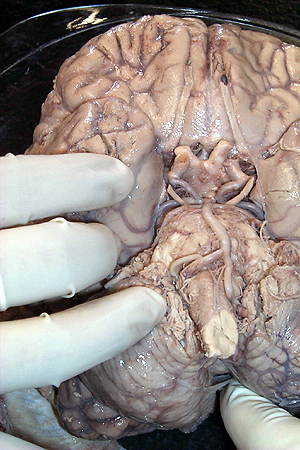
Hopefully, when you turn to the ventral surface of your brain specimen to look at vessels and nerves, it will resemble what you see in the picture below:
This specimen is fairly representive of what you will see in the lab. While not perfect, most of the major arteries and several nerves are well preserved. Your goal is to identify what you can, learn how to find what you do not immediately see and come to an understanding of the spatial relationships between nerves, arteries and external landmarks so as to be able to interpret what you will see in the clinic using imaging technology. On the next couple of pages, you'll see some hints on what to expect and how to go about looking for specific landmarks. |
| page1 |
|