![]()
| Introduction
Brainstem Landmarks |
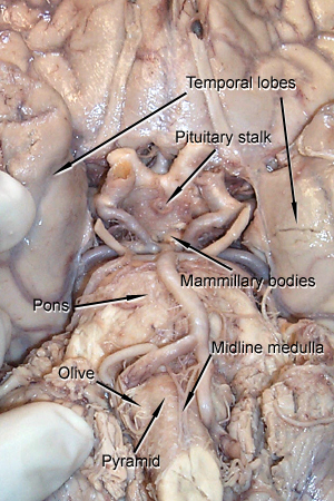
A close-up of the ventral surface of the brain is shown below. This specimen is especially good since the brainstem is intact and attached with the first cervical segment of the spinal cord. In most of your specimens, the brainstem cut will probably be much higher.
You should be able to easily identify the pons, the big bulbous mass of brainstem lying between the two temporal lobes of the cerebral hemisphere. Below the pons, you'll see the ventral surface of the tapering medulla oblongata. Just below the pons, identify the ventral medial sulcus in the midline. On each side of the sulcus, you should be able to identify two bumps. The most medial bumps are called the pyramids. The pyramids are comprised of axons of motor neurons that are found in the cerebral cortex and project all the way to the spinal cord. Just lateral to the pyramids, almost on the side of the medulla, you should find another bump called the olive. Later on, when we study the organization of the brainstem, you'll discover that this bump is formed by a squiggly-looking nucleus called the inferior olive. Neurons of the inferior olive project to the cerebellum and form one part of the circuitry involved in fine motor control. Rostral to the pons, between the two temporal lobes, we can just see the floor of the hypothalamus which is the ventral part of the diencephalon. We can see the stump of the pituitary stalk (the pituitary is left in the base of the skull in the sella turcica). Below that we can just see a pair of protuberances under the pons known as the mammillary bodies. These structures mark the caudal limits of the hypothalamus. You'll be able to use these structures as landmarks to locate the stumps of several of the cranial nerves that exit from the brainstem.
|
| page2 |
|