![]()
 |
Case
Study 1 -
|
| Imaging
arteries
Idenifying arteries normal carotid angiogram, lateral normal vertebral angiogram, lateral Veins and sinuses
|
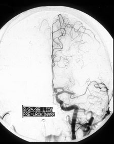
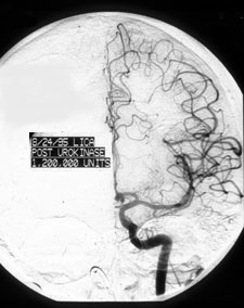
arteriesThe first cerebral angiogram you see below shows the result of an occlusion of a stem of the left middle cerebral artery (LMCA). Compare this image to the second one taken from the same patient that same day following anticogulant (urokinase) treatment.
Each major brain artery has a relatively consistent territory it supplies. Thus, occlusion of a brain blood vessel may result in tissue damage in a well demarcated zone. This is called an ischemic stroke. Stroke is recognized by a sudden loss of one or more neurological functions. |
| page1 |
|