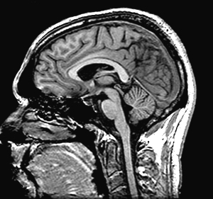
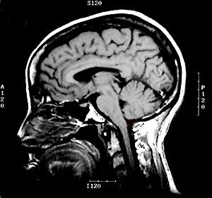
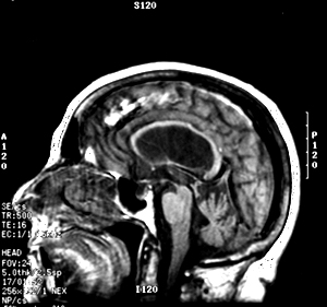
Once you
are finished with Neuroanatomy, the only brain slices you'll be
likely to see will be computerized images. Thus, structures
that are relatively easy(?!) to identify in your laboratory specimens
will have to be found again and again on film. After looking
at some of the cases presented here, you can imagine that doing
this is not always easy.
See if
you can identify the following structures on the following sagittal
MRIs from Cases A-C (normal, Alzheimer's and motor
neuron disease). Pass the cursor over the images at the points
which you think correspond to the various structures. Click on that
spot; if you're right, you'll be rewarded with the answer.
Remember, the dark areas on T1 weighted images represent fluid-filled
spaces like ventricles, canals and cisterns (don't forget that they
can also be blood vessels) and make for perfect landmarks in finding
your way around these brain images.
Question
- Find
the following landmarks:
-
Fourth Ventricle
-
Cerebral Aqueduct
-
Lateral Ventricle
-
Cisterna Magna
-
Foramen of Monro.


