![]()
| Introduction
Cases normal mri |
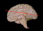
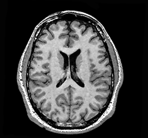
In infants, children and teenagers, the ventricles are normally small. Case A shown below is an MRI image of a normal brain. This is a horizontal section just below the corpus collosum.
Note the size of the lateral ventricles (if you're not sure what they are, they're the black "crescents" in the middle of the hemispheres) and the size and appearance of the gyri and sulci. Then, go on to the next case.
|
| page3 |