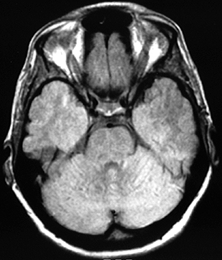
Horizontal MR Imagesintroduction | horizontal 1 | horizontal 2 | horizontal 3 Here's a section that you won't see in the lab, but you should recognize this slice representing the area between the two temporal lobes that you looked at in Laboratory 1 when you were studying the Circle of Willis. Try to identify the following areas (this is also a T2-weighted image): gyrus rectus, optic nerve, internal carotid artery, middle cerebral artery, basilar artery. Click on the image to see the labels. Other structures that you might be able to find include the ophthalmic artery, infundibulum and superior cerebellar artery.
Continue to sagittal sections ...
|