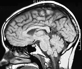
Sagittal MR Imagesintroduction | sagittal 1 | sagittal 2 Here's a classic mid-sagittal section. You've already studied this section in Case Study 2 with respect to identifying ventricles and cisterns (if you haven't viewed this case study, do so - it's very important!). Try to identify the following: fornix, optic chiasm, parieto-occipital sulcus, pituitary, superior colliculus. Click on the image to see the labels. What else can you identify?
|