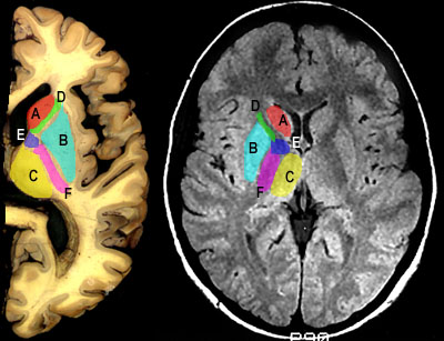
Horizontal MR Imagesintroduction | horizontal 1 | horizontal 2 | horizontal 3 Below you'll see a horizontal section through the internal capsule similar to the third section you made in the fourth lab, and its corresponding horizontal MR image. These horizontal images are T1/T2 hybrids; while white matter is darker than grey matter, ventricles are not enhanced. In the lab, the films that you will study will consist of pairs of horizontal sections. The first image at eacl level will be the hybrid image. The second will be the true T2 image. On one side of the section, you'll see that some areas are highlighted and labeled. Identify these areas. Click on the labeled area in the MR image to see the answer. Lateral to the putamen you can see the insular cortex. What do the small dark "holes" within this cortical area in the MRI represent?
|