|
Step 1 | Step 2 | Step 3 | Step 4 | Step 5 | Step 6 | thumbnails
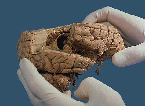
Figure 3. The cuts through the parietal and temporal lobes have been completed as described in the introduction, and the brain is being pulled apart. The "C" shaped fornix is seen rising out of the temporal lobe and then turning upward to become suspended from the corpus callosum. Click on the picture to see a closer view.
|