![]()
| Imaging
arteries
Idenifying arteries introduction normal carotid angiogram, lateral normal vertebral angiogram, lateral Veins and sinuses |
It is not necessary for the general physician to learn the cerebral vasculature in great detail, but a familiarity with the anatomy of key vessels, their distributions, and at some point clinical correlation of stroke in them, will be invaluable. As indicated in the laboratory excercise, some of the key arteries are:
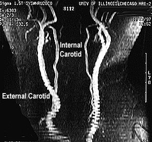
Picture studies specifically designed to show blood vessels are called angiograms. On the following pages, try to identify the blood vessels listed above on the pictured radiological studies. [Note: Not every vessel will appear in each study.] You will see both conventional angiograms (i.e. X-ray images obtained with dye injected into the vessel) and magnetic resonance angiograms (MRAs), images reconstructed from MRI flow signal voids created by blood moving within the MRI instrument. Hint: Refer first to your Neuroanatomy textbook or atlas before attempting to look at angiograms. It is a bit more difficult than you might think to identify vessels on angiograms. First, you must determine what is the view or perspective from which you are viewing the cerebrovascular tree. Standard views are AP (anterior-posterior) and lateral. Others views include oblique (non-orthogonal views). Second, you must not let the numerous overlapping vessel branches keep you from honing in on the vessel of interest. A key point to remember is that the internal carotid artery has NO branches in the neck and its first intracranial branch is the ophthalmic artery. The external carotid has numerous branches in the neck and head. This is shown in the MRA below which shows an anterior view of the carotids.
|
| page2 |