| Introduction
Cases
case
1
case
2
case
3
case
4
For
Discussion
quality
of life
timing
|
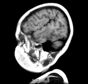
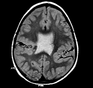
Images "A"
and "B" shown below were taken from the same individual.
MRI "A" is a sagittal section taken laterally through
the temporal lobe. "B" is a horizontal MRI scan.
Click on each image to see a comparable MRI scan from a normal inidividual.
A
B
Can you
identify the problem? Click on the icon to learn the diagnosis.
|