| Introduction
Cases
case
1
case
2
case
3
case
4
For
Discussion
quality
of life
timing
|
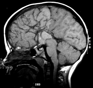
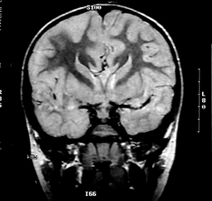
Images "A"
and "B" shown below were taken from the same individual.
MRI "A" is a standard midsagittal section. "B"
is a coronal MRI scan. "C" is a coronal scan taken from
a comparable level to that shown in "B" from a normal
individual.
A
B
C
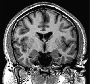
Can you
identify the differences? Click on the icon to learn the diagnosis.
|