Somatosensory 1;
Conscious Touch and Proprioception;
Unconscious Pathways
Competencies:
- Distinguish the receptors for touch, pressure and proprioception.
- Diagram the location of the axons, and synaptic way stations, for fine tactile discrimination.
- Relate the role of sensory information in cerebellar control of motor function.
- Compare the similarities and differences of dorsal root ganglia and trigeminal ganglion.
- Draw the organization of the cerebral cortex somatosensory homunculus.
To master
the material presented in this lecture:
Read ...
Purves text, Chapter 9
Haines
pp 188. 190.
Look at the Review Questions below ...
Listen
to the lecture and focus on the following points ...
- Different
receptor types can selectively respond to different types of energy,
and transduce the energy into nerve impulses (action potentials).
The brain keeps these signals about different stimuli segregated
for purposes of localization and interpretation. Much of neuroanatomy
has to do with learning the segregated pathways (or tracts).
- Sensory
receptors provide: localization of a stimulus, by virtue of their
place in the body.
- Specificity with regard to type of stimulus, by virtue of their structure.
- Intensity of stimulus information by virtue of the number of action
potentials/sec in the sensory axon.
- A
higher density of receptors gives finer spatial discrimination.
Thus, areas of the body with a high density of receptors will
need more processing neurons in the CNS. For example: A finger-tip,
high density, needs as many cerebral cortical neurons as the
larger shoulder which has a low density of receptors.
- Receptors
may be rapidly adapting, thus being useful for recognizing edges,
vibration, and direction of a moving stimulus on the skin. Examples
are: Pacinian corpuscle, Meissner's corpuscle and peritrichial
endings.
- Receptors
may be slowly adapting, thus being useful to recognize sustained
touch or stretching of the skin. Examples are: Merkel's disc and
Ruffini end organs.
- Receptors
in muscle, Proprioceptors, help you know "where you are in
space". These signal the onset of muscle contraction, the
degree of muscle shortening, and the stretch of muscle and tendon.
Examples are: Muscle spindles and the Golgi tendon organs.
- Receptors
respond to their preferred stimulus by causing, in the end of
the primary afferent axon, a receptor potential. This, depolarizing,
receptor potential is continuously graded, and the axon transforms
this signal into action potentials. The frequency of action potentials
is a measure of the intensity of the stimulus.
- The
primary afferent axon will carry the information about the sensory
stimulus towards the central nervous system. This axon has a cell
body, which is found in the dorsal root ganglion. Although developmentally,
these neurons went through a bipolar stage, they soon merged the
two poles, and are thus said to be unipolar (or pseudo-unipolar).
The peripheral process is the primary afferent axon in touch with
the receptor, and the central process of this neuron forms the
dorsal root, which enters the spinal cord.
- The
basic arrangement for the somatosensory pathways to conscious
(cortical) levels in the CNS is shown in the accompanying figure:

- The
primary afferent enters the CNS and has a synapse on the 2nd neuron.
- The
secondary neuron quickly sends its axon to the other side of CNS,
i.e. contralateral to the origin of the stimulus.
- The
tertiary neuron, in thalamus, sends its axon to cerebral cortex:
contralateral cortex analyzes the stimulus.
- The
larger axons in this figure represent the dorsal column - medial
lemniscus system. The axon enters at the dorsal lateral fissure
and joins the adjacent dorsal column. This system carries fine
tactile, discriminatory sensations and position sense(proprioception).
Subdivisions of the dorsal column are a medial fasciculus gracilis,
and a lateral fasciculus cuneatus. Fasciculus gracilis carries
information from below T6, i.e. principally lower extremity, and
F. cuneatus carries information from above T6.
- The
dorsal column axons end by a synapse in nucleus gracilis, or nucleus
cuneatus respectively, which are found in the medulla of the brain
stem.
- The
axons from neurons of n. gracilis and n. cuneatus cross to the
contralateral side, through internal arcuate fibers to form the
medial lemniscus. The segregation of the signals, i.e. somatotopic
arrangement of the fibers, continues with the upper extremity
being most dorsal in the tract, then body, lower extremity, and
most ventral the feet. One can imagine a (headless) person (or
homunculus) standing in the tract.
- The
medial lemniscus continues to ascend in the brain stem, changing
orientation in pons and midbrain while maintaining the somatotopic
segregation, and ends in a thalamic nucleus called Ventral posterior
lateral (VPL).
- Neurons
of the VPL project to primary somatosensory cortex, in the postcentral
gyrus of the parietal lobe. The homunculus is now oriented with
the feet on the medial surface of the hemisphere (in paracentral
lobule) and body, upper extremity and hand progressively represented
down the side of the hemisphere on the postcentral gyrus.
- UNCONSCIOUS "PROPRIOCEPTION":
- The sensory information discussed
above, which is useful for consciousness, is also useful for unconscious
functional control of movement. Therefore:
- The
primary afferent axon, upon entering the spinal cord, will have
branches (collaterals) to share the sensory signal with "local
reflex" neurons, and
- To share
the signal with the cerebellum (works at unconscious level) by
a synapse with spinal cord neurons, whose axons form spinocerebellar
tracts.
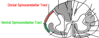
- Dorsal
spinocerebellar tract:
- Arises
from neurons in the nucleus dorsalis of Clarke and enters
the ipsilateral cerebellum via the inferior cerebellar peduncle.
- Ventral
spinocerebellar tract:
- Arises
from border cells of spinal gray and the axons ascend partly
crossed to enter the cerebellum via the superior cerebellar
peduncle. Most of the crossed fibers are thought to recross
once in the cerebellum.

- Cuneocerebellar
tract:
- Arises
from the external (accessory) cuneate nucleus and the axons
enter the ipsilateral cerebellum through the inferior cerebellar
peduncle.
- NOTE:
Cerebral cortex processes sensory information from the contralateral
side of the body, while cerebellar cortex processes sensory information
from the ipsilateral side of the body. Therefore, a lesion in
the left postcentral gyrus could give rise to sensory deficits
on which side? A lesion in the left cerebellum could give rise
to motor deficits on which side?
- The trigeminal system conveys sensations for the face similar to the spinothalamic and the dorsal column - medial lemniscus systems.
- The three divisions of the trigeminal nerves meet the sensory distribution from the dorsal roots of C2 and C3 from the crown of the head to the mandible and neck.
- Divisions of V:
- Ophthalamic = V1
- Maxillary = V2
- Mandibular = V3 (includes motor root)
- Trigeminal Ganglion (Gasserian, semilunar)
- Trigeminal nuclei and tracts: should be identified in each medullary and pontine section of the UIC Brainstem set
- Spinal tract and Nucleus will be discussed in the lecture on pain pathways
- Trigeminothalamic tract, contralateral to sensory origin and ends in VPM thalamus.
- Main Sensory Nucleus (Principal or Chief), relays discriminating tactile sensation of face
- Mesencephalic tract and nucleus, unique unipolar cells innervating stretch receptors such as the muscles of mastication.
- Motor nucleus of V.
- Receives bilateral cortical (corticobulbar) input.
- Trigeminal reflexes can test if nerves are intact.
- Jaw jerk reflex, tests afferent limb on V3 and synapse at trigeminal motor nucleus, and the efferent limb on V3
- Corneal reflex, tests afferent limb on V1 and synapse at facial motor nucleus, and the efferent limb of facial nerve.
Consider the Following Questions ...
- Where is the cell body of a sensory neuron with connection to intrafusal muscle fibers in the biceps?
- Describe the places in spinal cord this neuron will send collateral branches and terminals.
- What does it mean that a receptor is a "transducer" and that it responds best to a "particular stimulus"? Describe some receptors for: a) muscle stretch; b) touch; c) pain.
- Describe the somatotopic organization of the postcentral gyrus, and identify which part of the internal capsule contains these thalamic radiations.
|