Organization of the Brainstem and Reticular Formation
Competencies:
- In the brain stem, distinguish the locations of basilar, tegmentum and tectum components.
- Diagram a cross section of midbrain and locations of major tracts and nuclei.
- Construct a suprasegmental integrative system in the brain stem.
- Appraise the locations and connections of the monoamine cell groups in brain stem.
To master
the material presented in this lecture:
Read ...
Purves text, pg 720-728, 390-392.
Look at the Review Questions below ...
Listen
to the lecture and focus on the following points ...
 The
Brainstem and Reticular Formation The
Brainstem and Reticular Formation
- The
BRAIN STEM provides the continuity of connections from the
spinal cord to the higher CNS. It is located in the posterior
cranial fossa along with the cerebellum.
- CRANIAL
NERVES connected to the medulla are #6 through #12. Therefore,
much of the muscle control of, and sensations received from the
head relate to the medulla, and in this way, the brain stem is
similar to the spinal cord.
- Brain
stem differs from spinal cord.
- connections
to the cerebellum
- vital
centers', in its core, e.g. cardiovascular and respiratory
centers.
- 'open'
ventricular system.
- special
sensory afferents (e.g. hearing) and special motor efferents
(e.g. muscles of facial expression).
- basilar
myelinated tracts;
- cerebral
peduncle of midbrain
- basilar
pons
- pyramids
of medulla.
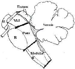
- Learn
the external features of the brain stem, which you will see again
in the cross sections
- identify
the limits of the Medulla, the Pons and the Midbrain
- how
do the fourth ventricle and cerebral aqueduct relate to the
different levels?
- Vascular
accidents or tumors in or on the brain stem give wide spread clinical
deficits, because of the long tracts found here, therefore, cranial
nerve signs will usually be the best localizing indicators.
- The Pons has nuclei for cranial nerves #5 to #8.
- The vital centers for respiratory and cardiovascular control are at junction with medulla.
- The Pons is connected to the cerebellum from the basilar nuclei through the middle cerebellar peduncle.
- The trigeminal nerve enters through middle cerebellar peduncle to reach its nuclei found throughout the brain stem.
- The fourth ventricle is widest at the junction of pons and medulla.
- The Midbrain has cranial nerves #3 and #4 which help move the eyes.
- Midbrain superior colliculi modify visual reflexes.
- Midbrain inferior colliculi relay auditory sensations to thalamus.
- Tectum refers to the superior and inferior colliculi collectively.
- Tegmentum is found throughout the brain stem, below the ventricular system.
- Basilar structures on the ventral side of brain stem are mostly myelinated fiber tracts.
- The Reticular formation is found in the tegmentum of brain stem.
- Certain cell groups may be stained to show particular transmitters (NE, E, DA, 5HT, ACh). Like other neurons of reticular formation these cells have long axons.
- Reticular neurons receive information from several sensory sources, and integrate appropriate responses for the animal, e.g. respiration, autonomic drive on viscera, upright posture, and levels of alertness of the cerebral cortex.
- Reticular formation is a suprasegmental integrative area.
Consider the Following Questions ...
-
Describe the organizational pattern of the motor nuclei vs the sensory nuclei within the brain stem. The tectum, tegmentum and basal portions refer to what regions of the mesencephalon?
-
As it grows larger and larger, an auditory nerve tumor in the ponto-medullary junction, would progressively affect what other cranial nerves?
-
The neurons of the raphe nuclei and locus coeruleus use which monoamine transmitters?
-
Compromising the function of the reticular formation in the midbrain (e.g. herniation of the temporal lobe into the tentorial notch) might result in a patient (1) constantly awake or (2) asleep? What is the ascending reticular activating system?
.
|