|
Gross Organization of the CNS and Spinal Cord;
Blood Supply and Ventricles
Competencies (Gross Organization):
- Diagram components of the CNS and PNS.
- Differentiate the lobes of cerebral hemisphere.
- Illustrate the cross sectional anatomy of the spinal cord.
- Assess the relationship of bone, meninges and CSF to the CNS.
Competencies (Blood Supply and Ventricles):
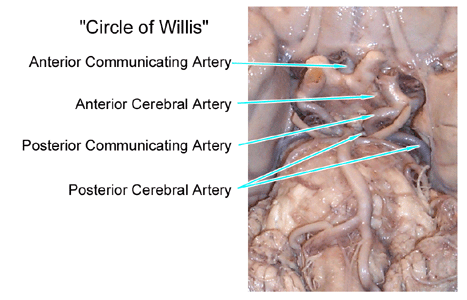
- Diagram the arterial circle of Willis and its relationships on the base of the brain.
- Predict the regions of supply of the anterior, middle and poster cerebral arteries.
- Distinguish the Blood Brain Barrier from that found in e.g. liver.
- Appraise the points in CSF flow where obstruction is likely to occur.
To master
the material presented in this lecture:
Read ...
Purves text, pp 717-720, 735-744.
Look at the Review Questions below ...
Listen
to the lecture and focus on the following points ...
- Dermatomes and myotomes of the the PNS have specific patterns of innervation.
- Afferent axons carry sensory information to the CNS.
- Efferent axons carry motor information to muscles or glands.
- Autonomic innervation is craniosacral/parasympathetic or thoracolumbar/sympathetic.
- Paired arteries rise through the neck to supply the brain: internal carotid arteries and vertebral arteries.
- Meninges (pia, arachnoid and dura), and fluid (CSF), and bones protect the brain.
- Spinal cord is the segmented part of CNS.
- CNS in the head is supra-segmental.
- Posterior cranial fossa houses brain stem, cerebellum and 10 pairs of cranial nerves.
- Above brain stem and above tentorium cerebelli is the supra-tentorial compartment.
- Supra-tentorial compartment houses diencephalon and telencephalon (including basal ganglia) and 2 pairs of cranial nerves (1. olfactory, 2. optic).
- Cerebral hemispheres have fairly regular appearances allowing discussion of the localization of functions in different lobes: frontal, parietal, occipital and temporal lobes, (what bones overlay these lobes?).
- Another lobe at the medial edge of the hemisphere is the “limbic lobe” often studied in physiological psychology courses.
- The spinal cord is the part of your CNS which resides in the vertebral column. While diminutive in size, the spinal cord is the principle source of control of the muscles of your body, and the entry point of sensations from the body.
- A ‘cervical enlargement' and a ‘lumbo-sacral enlargement' of the spinal cord, reflect the need for more neurons at those levels to control the large extremities.
- Internal spinal cord, in cross sections:
- Dorsal roots (sensory, afferent) enter at the dorsal lateral sulcus
- Ventral roots (motor, efferent) leave at the ventral lateral sulcus.
- White matter (myelinated axons) around the outside (increases as ascend cord).
- dorsal funiculus (dorsal column)
- Lissauer's tract
- lateral funiculus
- fasciculus proprius (next to gray matter, connections stay within sp cd)
- ventral funiculus
- Gray matter (mainly neurons and their dendrites)
- dorsal horn
- intermediate gray
- ventral horn
- Nuclear organization and Rexed Laminae
- posteromarginal nucleus = Rexed Lamina I (mostly pain)
- substantia gelatinosa = Rexed lamina II (modulation of pain)
- nucleus proprius (principle) = Rexed laminae III - V (mixed sensory)
- intermediate gray = Rexed laminae VI and VII (integrative functions)
- lateral motor columns = Rexed lamina IX (lower motor neurons for leg and arm)
- medial motor columns = Rexed lamina IX (lower motor neurons for axial, i.e. trunk muscles)
- With a tumor or vascular accident the highest segmental problems usually indicate where the problem occurred.
- Strokes:
Usually sudden onset.
- Brain
has a high metabolic rate, and does not store oxygen or glucose.
- Vascular
insufficiency results in ischemia and continued lack of nutrients
results in infarct.
- Constant
blood flow over a wide range of Blood Pressure.
- Vascular
innervation and control: appears to be mostly local control
via concentration of metabolites from neuronal and glial activity.
- Collateral
circulation via anastomosis at the arteriolar level. There are
few end arteries.
- Circle
of Willis:
- Internal carotid arteries with anterior cerebral arteries, anterior communicating artery, posterior communicating arteries and posterior cerebral arteries.

- Internal
Carotid Artery System. (Anterior Circulation)
- Lenticulostriate
(lateral striate A's) - A's of cerebral hemorrhage.
- Vertebral-Basilar
System. (Posterior Circulation)
- To brainstem, cerebellum and posterior cerebral A's
- Venous
drainage: no valves
- dural sinuses
- emissary veins, infections from
scalp.
- Blood
Brain Barrier: (BBB) Endothelium of brain vessels.
- Cerebrospinal
Fluid: (CSF)
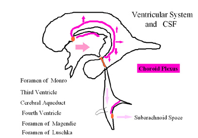
- Choroid
Plexus epithelium: barrier and secretory.

- Flow
route may be obstructed at many sites and cause hydrocephalus.
- Hydrocephalus may be "communicating" or "non-communicating" - define!
- Lateral
ventricles (1 & 2)
 foramina of Monro (1&2) foramina of Monro (1&2)  3rd
ventricle 3rd
ventricle  cerebral aqueduct cerebral aqueduct  4th ventricle 4th ventricle medial
and lateral foramina medial
and lateral foramina  subarachnoid space subarachnoid space  cisterns cisterns  arachnoid villi arachnoid villi  veinous system veinous system
Consider the Following Questions ...
-
The delicate central nervous system is protected by three membranes. Name the three membranes and describe their relationship to each other. Which two are directly exposed to the CSF, and how is the CSF moved to the venous blood?
-
What parts of the CNS are found in 1,2,3, below, and in which of these areas on the left would you look for the origin of the clinical problems labeled a, b, c?
| (1) in the posterior cranial fossa? |
(a) a T-10 segmental problem? |
| (2) above the tentorium cerebelli? |
(b) a facial nerve paralysis? |
| (3) in the vertebral column? |
(c) an inability to name something that is seen? |
- Describe the organizational pattern of the motor nuclei vs the sensory nuclei within the brain stem.The tectum, tegmentum and basal portions refer to what regions of the mesencephalon?
- Groups of axons with a similar function travel together in a tract or fasciculus, and they will often be named for their sites of origin and of termination. For example, where would the Corticospinal tract begin and end? How about the Spinocerebellar tract?
- The spinal cord is organized such that the cells (gray matter) are located where? And where are the long myelinated axons found?
- The cervical enlargement of the spinal cord is found at what level, and why is it enlarged?
- Which nuclear groups particularly characterize the T1 to L2 spinal cord gray matter?
- Draw and label the circle of Willis, and determine the area of cerebral cortex that will be affected by occlusive disease of the Anterior Cerebral Artery.
- What cellular specializations of choroid plexus contribute to making CSF a part of the relatively privileged environment of the CNS?
-
Name the spaces CSF traverses from lateral ventricles to the superior sagittal sinus, and identify the spaces that will be enlarged if a disease process closes the cerebral aqueduct.
|