![]()
| Introduction
Ventral Surface
|
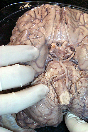
You have already found some of the prominent landmarks on the ventral surface of the brain in the first laboratory session. Take a look at the image below and see if you now recognize features such as the olfactory tract, pituitary stalk and the uncus, the prominent "hooking" cortex on the medial surface of the temporal lobe just lateral to the carotid artery (it might be tricky to see since the meninges are still intact in this image. Find the gyrus rectus and the orbital gyrus on the ventral surface of the prefrontal cortex. Why do you think it's called the orbital gyrus? See if you can also find the flocculus at the base of the cerebellum just lateral to the pontine medullary juction. Move the cursor over the image to see the labels.
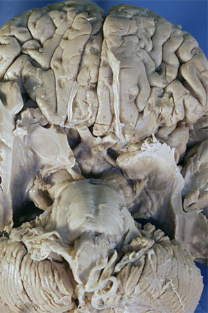
The next image takes a slightly different view of the ventral surface. Meninges and blood vessels have been removed and the temporal lobe has been sectioned through the ventricle and the base of the temporal lobe removed. We will study this prosection in more detail in Laboratory 6. In the meantime, you now should be able to follow the olfactory tract as it "disappears" toward the base of the forebrain (hence, the basal forebrain). This is where you will find the anterior perforated substance. Why is the substance perforated? You should easily find the uncus on the medial surface of the temporal lobe pointing toward the pituitary stalk. By the way, what is that "almond" shaped nucleus that has been revealed lying within the uncus? Posterior to the pituitary stalk you should now easily see the mammillary bodies, which mark the posterior edge of the diencephalon. Behind that you will find the interpeduncular fossa, the "space" (inter-) between the two cerebral peduncles. You can at least see one nerve coming from the interpeduncular fossa. What is that nerve? Move the cursor over the image to see the labels.
|
| page4 |